Veterinary Dental X-Ray Positioning: A Comprehensive Guide
Understanding precise techniques, like parallel and bisecting angles, is crucial for optimal imaging of canine and feline dentition, as detailed in educational resources.
Veterinary dental radiology is a cornerstone of modern veterinary practice, enabling the non-invasive assessment of both visible and hidden dental structures. Accurate radiographic interpretation relies heavily on proper x-ray positioning, a skill honed through dedicated study and practical application. Resources like Beyond The Crown Veterinary Education emphasize the importance of mastering techniques such as the parallel and bisecting angle methods.
This guide provides a comprehensive overview of these techniques, focusing on maximizing diagnostic quality while minimizing radiation exposure. Understanding tube head angulation, patient positioning, and potential artifacts is paramount. The field is also evolving with advancements like AI-powered image analysis, exemplified by RapidRead Dental, enhancing diagnostic capabilities and efficiency for veterinary professionals.
Importance of Accurate Positioning
Accurate radiographic positioning is absolutely critical in veterinary dentistry. Improper angles or beam alignment lead to geometric distortion, resulting in misdiagnosis and potentially inappropriate treatment plans. Overlapping structures, foreshortening, or elongation of tooth roots can obscure pathology like fractures, root canal issues, or periodontal disease.
Resources emphasize positioning the patient correctly, ensuring the area of interest is closest to the x-ray beam. Mastery of parallel and bisecting angle techniques, as detailed in iM3 positioning kits and educational materials, is essential. Consistent, precise technique minimizes retakes, reducing radiation exposure and improving diagnostic confidence. Ultimately, accurate positioning directly impacts patient care and welfare.
Radiographic Techniques in Veterinary Dentistry
Two primary techniques dominate veterinary dental radiology: the parallel technique and the bisecting angle technique. The parallel technique, ideal for caudal mandibular premolars and molars, requires the x-ray beam to be parallel to the tooth root, minimizing distortion. The bisecting angle technique, used for incisors, canines, and sometimes premolars, aims the beam perpendicular to an imaginary line bisecting the angle formed by the tooth and the film.
Proper angulation, as highlighted in resources like Beyond The Crown, is vital. Tube head positioning – typically 45-55 degrees for maxillary molars – influences image quality. Understanding these techniques, alongside utilizing guides like those from iM3, ensures comprehensive radiographic evaluation and accurate diagnoses.
Equipment and Preparation
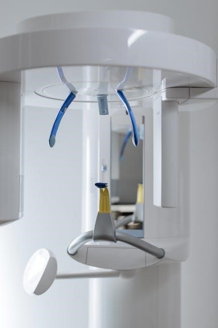
Essential equipment includes dental x-ray units, either digital or film-based, alongside positioning aids like bisecting angle guides for accurate image capture.
Dental X-Ray Units: Types and Features
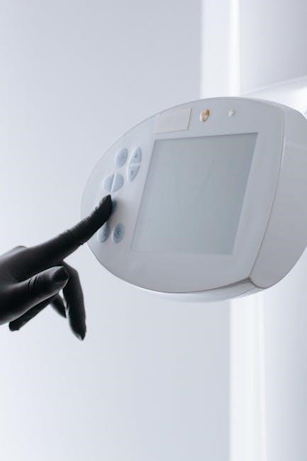
Veterinary dental radiology relies on specialized x-ray units designed for the smaller oral structures of animals. These units generally fall into two categories: wall-mounted and portable. Wall-mounted units offer consistent power and geometry, ideal for high-volume practices. Portable units provide flexibility, allowing imaging in various locations, even during anesthesia.
Key features include adjustable kVp and mA settings to control penetration and contrast, crucial for different tooth sizes and densities. Tube head design impacts beam collimation, minimizing scatter radiation. Digital units offer immediate image viewing and manipulation, while film-based systems require processing. Modern units often incorporate timers for precise exposure control, enhancing image quality and patient safety.
Digital vs. Film-Based Radiography
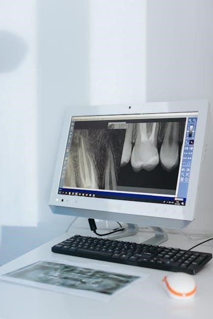
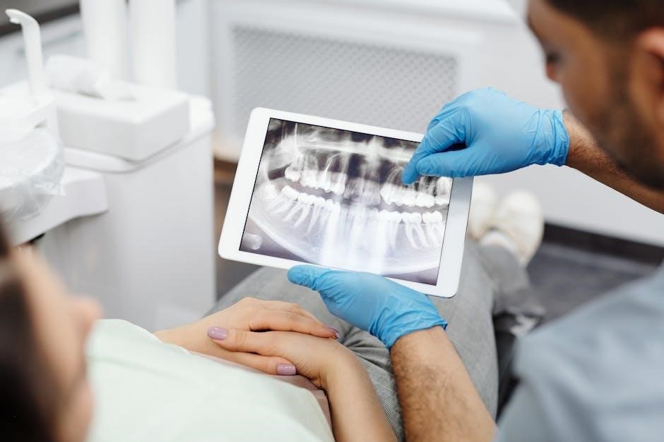
Digital radiography is rapidly becoming the standard in veterinary dentistry, offering significant advantages over traditional film-based systems. Digital sensors capture images instantly, eliminating the need for chemical processing and reducing exposure time. This allows for immediate image review, enhancing diagnostic efficiency and patient care. Image manipulation, such as adjusting brightness and contrast, is easily achieved digitally.
Film-based radiography, while less common now, remains a viable option. It requires careful processing to develop the latent image, and quality control is paramount to avoid artifacts. Digital systems, like those incorporating AI (RapidRead Dental), further improve image quality and diagnostic accuracy, representing a substantial advancement in veterinary dental imaging.
Patient Preparation and Safety Protocols
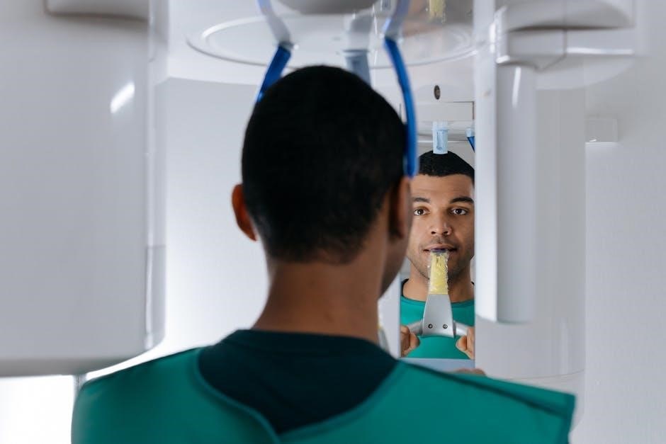
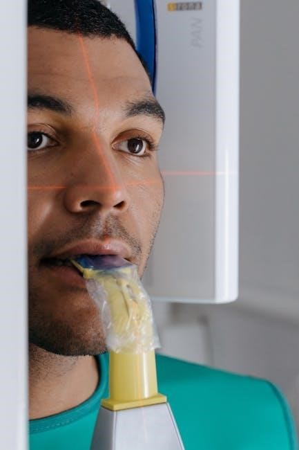
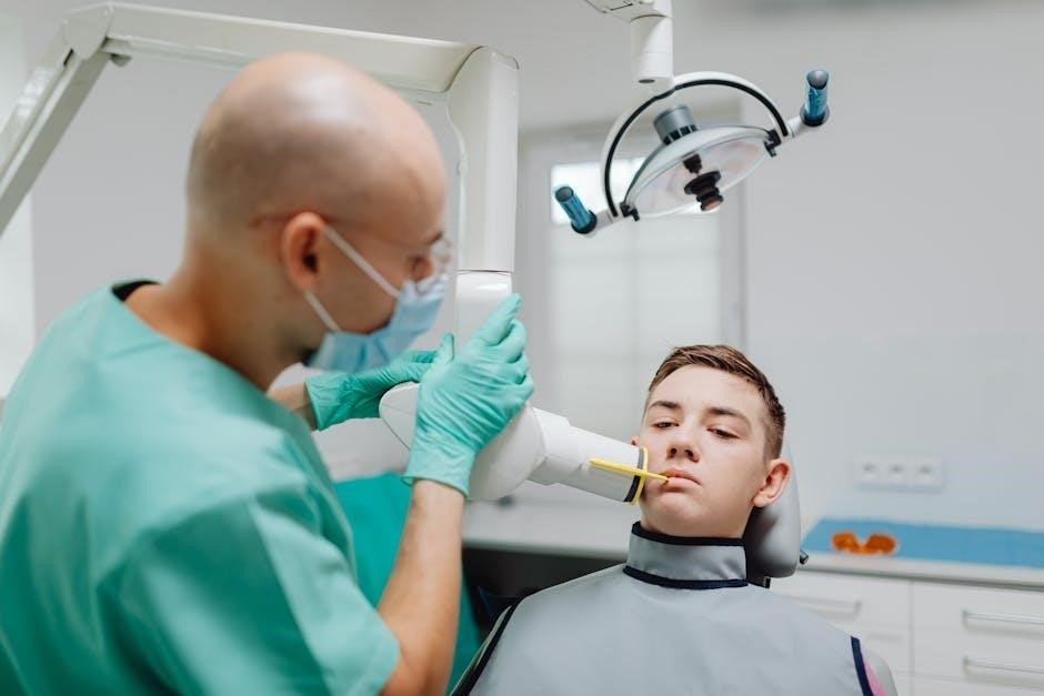
Prioritizing safety is paramount when performing veterinary dental radiography. Patient positioning is key – ensuring the area being imaged is closest to the radiographic beam minimizes exposure. Anesthesia is often necessary for optimal positioning and to prevent patient movement, ensuring sharp images.
Radiation safety protocols must be strictly adhered to, including the use of lead aprons for personnel and thyroid shields for the patient. Proper collimation limits the beam size to the area of interest, reducing unnecessary radiation. Regular quality control checks of the X-ray unit are essential, alongside meticulous record-keeping of radiation doses received by each animal.
Positioning Techniques: Maxilla
Maxillary imaging utilizes both parallel and bisecting angle techniques; angles of 45-55 degrees are often employed for optimal visualization of structures.
Maxillary Premolar and Molar Positioning (Parallel Technique)
Achieving accurate maxillary premolar and molar radiographs demands meticulous application of the parallel technique. This method requires the X-ray beam to be directed perpendicular to the tooth and the image receptor. Proper patient positioning is paramount, ensuring the area being imaged is closest to the beam.
Tube head angulation, typically between 45-55 degrees, is crucial for visualizing these posterior teeth effectively. Utilizing a bisecting angle guide can aid in achieving the correct angulation. The parallel technique minimizes distortion, providing a more accurate representation of the tooth’s anatomy, including the roots and surrounding bone structure. Consistent practice and attention to detail are essential for successful implementation.
Maxillary Incisor and Canine Positioning (Bisecting Angle Technique)
The bisecting angle technique proves invaluable for imaging maxillary incisors and canines, particularly when anatomical limitations hinder parallel positioning. This technique involves angling the X-ray beam midway between the tooth and the image receptor. Accurate angulation is vital; resources suggest aiming high and through the eye for optimal results.
Employing a bisecting angle guide with clear instructions significantly enhances precision. While potentially introducing some distortion, careful technique minimizes this effect. Proper patient positioning, bringing the target area close to the beam, remains crucial. Mastering this technique requires dedicated practice and a thorough understanding of dental anatomy.
Tube Head Angulation for Maxillary Teeth
Achieving correct tube head angulation is paramount for clear maxillary dental radiographs. For molars (110-109 & 210-209), a 45-55 degree angle is generally recommended, aiming high and through the eye, as highlighted in veterinary dental education materials. This angle facilitates visualization of the entire tooth root and surrounding structures.
Precise angulation minimizes distortion and superimposition of anatomical features. While the bisecting angle technique introduces some geometric distortion, careful alignment reduces its impact. Consistent practice and utilization of positioning aids, like X-ray bisecting angle guides, are essential for mastering this skill. Remember, optimal image quality relies on meticulous attention to detail.
Positioning Techniques: Mandible
Mandibular imaging commonly employs parallel and bisecting angle techniques, focusing on accurate beam alignment and patient positioning for diagnostic quality radiographs.
Mandibular Premolar and Molar Positioning (Parallel Technique)
The parallel technique for mandibular premolars and molars demands meticulous attention to detail. Positioning requires the X-ray beam to be directed perpendicular to the tooth and the image receptor, ensuring minimal distortion. This is achieved by aligning the beam parallel to the long axis of the tooth.
Proper patient positioning is vital; the animal’s head should be stabilized, and the mandible gently compressed to facilitate accurate film placement. Utilizing X-Ray Bisecting Angle Guides, available in complete kits like iM3’s, can significantly aid in achieving parallelism. Careful angulation is key to visualizing the entire tooth, including the apex and surrounding bone structure.
Consistent practice and hands-on training, such as those offered by Beyond The Crown Veterinary Education, are essential for mastering this technique and minimizing errors.
Mandibular Incisor and Canine Positioning (Bisecting Angle Technique)
The bisecting angle technique is frequently employed for mandibular incisors and canines due to anatomical limitations. Positioning involves directing the X-ray beam perpendicular to an imaginary line bisecting the angle formed by the tooth’s long axis and the image receptor. This method requires careful estimation of the bisecting angle to minimize distortion.
Utilizing X-Ray Bisecting Angle Guides, readily available from suppliers like iM3, greatly improves accuracy. Proper patient stabilization and gentle mouth opening are crucial. Tube head angulation is vital; typically, a 45-55 degree angle is recommended, though adjustments may be needed based on individual anatomy.
Consistent application of this technique, coupled with ongoing education, ensures diagnostic quality radiographs. Remember, mastering this requires practice and attention to detail.
Tube Head Angulation for Mandibular Teeth
Precise tube head angulation is paramount for clear mandibular radiographs. For premolars and molars utilizing the parallel technique, the beam should be directed perpendicular to the image receptor and tooth long axis. However, the bisecting angle technique, common for incisors and canines, necessitates angling the tube head.
Generally, a 45-55 degree angle is a starting point, but anatomical variations demand adjustments. Positioning the patient so the area being imaged is closest to the beam is key. Incorrect angulation leads to distortion – foreshortening or elongation – compromising diagnostic value.
Resources like Beyond The Crown Veterinary Education emphasize careful observation and adjustment. Consistent practice and utilizing positioning aids enhance accuracy, ensuring optimal image quality for effective dental assessment.
Specific Tooth Positioning
Individual tooth evaluation requires tailored techniques; fractured teeth present unique challenges, and root canal assessments demand precise positioning for optimal visualization.
Positioning for Individual Tooth Evaluation
When focusing on a single tooth, meticulous attention to radiographic principles is paramount. Utilizing both parallel and bisecting angle techniques allows for comprehensive assessment. The tube head angle must be adjusted to minimize distortion and ensure accurate representation of the tooth’s anatomy, including the apex and surrounding structures.
Careful patient positioning is also vital, bringing the area of interest as close as possible to the radiographic beam. This minimizes magnification and enhances image clarity. For isolated teeth, slight adjustments to the beam’s direction may be necessary to avoid superimposition from adjacent structures, providing a clear diagnostic view.
Proper restraint and stabilization are essential for obtaining high-quality images, especially when dealing with cooperative challenges.
Challenges in Positioning Fractured Teeth
Fractured teeth present unique radiographic challenges due to potential displacement of fragments and difficulty in visualizing the fracture line. Precise angulation is critical; slight adjustments to the tube head may be needed to reveal hidden fracture planes. Superimposition of adjacent structures can obscure the fracture, necessitating multiple projections from different angles.
Careful consideration must be given to patient positioning to minimize movement and ensure the fractured segments remain stable during exposure. Utilizing a fast shutter speed can reduce motion blur. Digital radiography offers advantages in these cases, allowing for image manipulation and enhancement to better delineate the fracture.
Accurate interpretation requires a thorough understanding of dental anatomy and fracture patterns.
Positioning for Root Canal Therapy Assessment
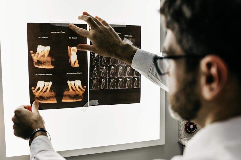
Evaluating teeth for root canal therapy demands detailed radiographic assessment of the pulp cavity, root canal length, and periapical structures. Parallel technique is generally preferred for accurate length determination, minimizing distortion. Multiple projections – at least two, ideally more – are essential to visualize the entire root structure in both dimensions.
Angulation should be carefully controlled to avoid superimposition of anatomical structures. Digital radiography allows for precise measurements of root canal length and width. Assessing for existing periapical lesions or evidence of previous treatment is crucial.
Proper positioning ensures accurate diagnosis and successful treatment planning for endodontic procedures.
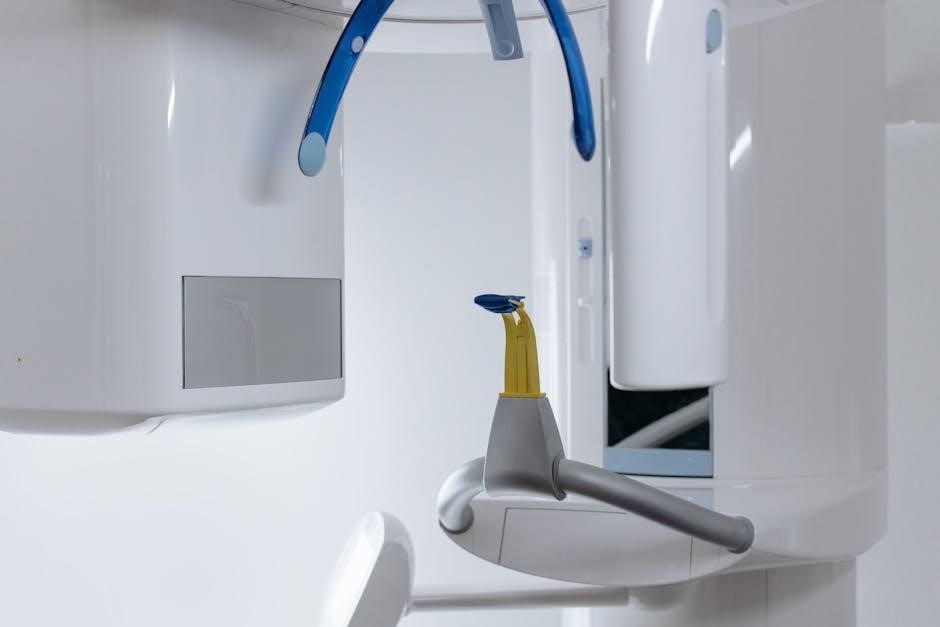
Image Evaluation and Artifacts
Accurate interpretation requires recognizing common artifacts and understanding their causes, ensuring reliable assessment of dental structures for diagnosis and treatment planning.
Recognizing Common Radiographic Artifacts
Radiographic artifacts can significantly compromise the diagnostic quality of dental x-rays. Motion blur, often appearing as streaks, results from patient movement during exposure. Positioning errors, like incorrect tube head angulation, lead to distortion, affecting anatomical representation.
Foreign objects, such as hair or metallic materials, create radiopaque shadows. Scatter radiation, reducing image contrast, arises from insufficient collimation or inadequate shielding. Film-based radiography is susceptible to processing errors – under or overdevelopment, or chemical contamination – causing density variations.
Digital systems can exhibit artifacts like pixelation or banding. Recognizing these artifacts is vital for differentiating them from actual dental pathology, ensuring accurate diagnoses and appropriate treatment plans. Careful technique and quality control minimize their occurrence.
Interpreting Dental X-Ray Images
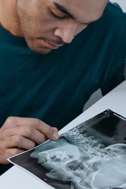
Accurate interpretation of veterinary dental radiographs requires a systematic approach. Begin by evaluating the periodontal ligament space – widening suggests periodontal disease. Assess alveolar bone levels; loss indicates bone resorption. Examine tooth roots for signs of fractures, root canal pathology, or periapical lesions (radiolucencies).
Evaluate the pulp chamber size and shape, noting any abnormalities. Look for evidence of resorptive lesions, often appearing as subtle radiolucent defects. Compare both sides of the mouth for symmetry.
Consider the patient’s clinical signs and a thorough oral examination alongside radiographic findings. Understanding normal anatomical structures and common pathologies is crucial for accurate diagnosis and treatment planning.
Quality Control and Image Optimization
Consistent quality control is paramount in veterinary dental radiology. Regularly assess radiographic technique – proper angulation, exposure settings, and film processing (or digital sensor calibration) are vital. Utilize test objects to evaluate spatial resolution and contrast. Implement a schedule for equipment maintenance and calibration.
Optimize image quality by minimizing motion blur through proper restraint and exposure time. Ensure adequate penetration to visualize all dental structures clearly.
Review images for artifacts and repeat exposures if necessary. Digital radiography offers advantages in image manipulation and optimization, but proper settings are still crucial. Documentation of quality control procedures is essential for maintaining high standards.
Advanced Technologies & Resources
AI, like RapidRead Dental, enhances diagnostic speed and accuracy, while iM3 kits provide comprehensive positioning tools for veterinary dental radiology workflows.
AI in Veterinary Dental Radiology (RapidRead Dental)
RapidRead Dental, launched by ANTECH in May 2025, represents a significant leap forward in veterinary dental diagnostics. This Artificial Intelligence (AI) powered technology is designed to assist veterinarians in interpreting dental radiographs with increased speed and accuracy. By automating aspects of image analysis, RapidRead Dental can help identify subtle anomalies often missed during manual review.
The integration of AI into dental radiology streamlines workflows, potentially reducing diagnostic time and improving patient care. While not replacing the expertise of the veterinary radiologist, it serves as a powerful tool to enhance their capabilities. Further research and adoption of such technologies promise to revolutionize the field, offering more efficient and reliable diagnostic outcomes for animal patients.
iM3 Complete X-Ray Positioning Kits
iM3 offers comprehensive X-Ray Positioning Kits specifically designed for veterinary dental use. These kits, intended for trained professionals, include essential tools to ensure accurate and consistent radiographic technique. Components often feature X-Ray Bisecting Angle Guides with detailed instructions, aiding in proper tube head alignment and image acquisition.
The availability of these kits simplifies the positioning process, promoting standardized protocols within veterinary practices. Decreasing or increasing quantities of individual components allows for customization based on specific needs. iM3 dental products are intended for use by veterinary personnel with appropriate training, supporting high-quality dental imaging and diagnosis.
Continuing Education and Hands-on Training
Investing in further training, particularly through hands-on laboratories, is highly recommended to accelerate the learning curve in veterinary dental radiology. Mastering radiographic positioning requires practical application and refinement of techniques. Educational resources emphasize the importance of positioning the patient strategically, ensuring the area of focus is closest to the radiographic beam for optimal image clarity.
These courses build upon foundational knowledge, addressing challenges like positioning fractured teeth and preparing for root canal therapy assessments. Consistent updates and participation in continuing education are vital for staying current with advancements and best practices in the field, ultimately improving diagnostic accuracy.
Legal and Ethical Considerations
Adhering to radiation safety regulations and maintaining meticulous record keeping are paramount for ethical and legally compliant veterinary dental radiology practices.
Radiation Safety Regulations
Compliance with federal and state regulations regarding radiation exposure is non-negotiable in veterinary dentistry. These regulations dictate permissible exposure limits for both personnel and patients, necessitating stringent safety protocols. Proper shielding – including lead aprons, thyroid shields, and protective eyewear – is essential for all personnel involved in radiographic procedures.
Regular calibration and maintenance of dental X-ray units are crucial to minimize radiation output while maintaining image quality. Detailed records of radiation exposure, equipment maintenance, and personnel training must be meticulously maintained for inspection. Furthermore, understanding the ALARA principle – “As Low As Reasonably Achievable” – guides practitioners to minimize radiation dosage whenever possible, prioritizing patient and staff safety.
Record Keeping and Documentation
Meticulous documentation is paramount in veterinary dental radiology. Each radiographic procedure requires a comprehensive record, including patient identification, date and time of exposure, specific teeth imaged, radiographic technique utilized (parallel or bisecting angle), and any observed artifacts. Detailed notes regarding patient positioning and tube head angulation are also essential for accurate interpretation and future reference.
Proper storage of both digital and film-based radiographs is critical for legal and diagnostic purposes. Maintaining a clear chain of custody for all images ensures accountability and prevents loss or misinterpretation. Accurate records facilitate effective communication with colleagues, aid in tracking dental health over time, and provide valuable documentation in the event of legal scrutiny.