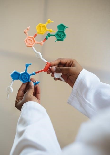
Genetic processes‚ including replication‚ transcription‚ and translation‚ are fundamental to life‚ building proteins from DNA and RNA instructions – a core concept for study.
Overview of Genetic Material
Genetic material‚ at its core‚ dictates the traits of all living organisms. DNA (deoxyribonucleic acid) serves as the long-term storage of this information‚ a stable blueprint passed down through generations. However‚ DNA doesn’t directly build proteins; it requires an intermediary. This is where RNA (ribonucleic acid) steps in‚ acting as a messenger carrying instructions from the DNA in the nucleus to the ribosomes in the cytoplasm.
The process of converting DNA into proteins involves two key steps: transcription and translation. Transcription creates mRNA from a DNA template‚ while translation uses mRNA to assemble amino acids into proteins; Understanding these molecules and their roles is crucial for grasping the central dogma of molecular biology and the very essence of heredity.
The Central Dogma of Molecular Biology
The central dogma describes the flow of genetic information within a biological system. Initially proposed by Francis Crick‚ it outlines the process from DNA to RNA to protein. Information travels unidirectionally: DNA is transcribed into RNA‚ and RNA is then translated into protein. While exceptions like reverse transcription exist‚ this remains a foundational principle.
Transcription‚ utilizing RNA polymerase‚ creates mRNA from a DNA template. Subsequently‚ translation‚ occurring at ribosomes‚ decodes mRNA into a specific amino acid sequence‚ forming a polypeptide chain – the building block of proteins. This dogma highlights how genetic instructions are expressed‚ driving cellular function and ultimately‚ life itself.

DNA Structure and Function
Deoxyribonucleic acid (DNA) stores genetic information‚ dictating traits and enabling heredity through its unique double helix structure and nucleotide composition.
Double Helix Structure of DNA
The iconic double helix‚ first elucidated by Watson and Crick‚ describes DNA’s structure – two strands intertwined around a central axis. These strands are antiparallel‚ running in opposite directions‚ and held together by hydrogen bonds between complementary nitrogenous bases. Adenine (A) always pairs with Thymine (T)‚ while Guanine (G) pairs with Cytosine (C). This specific pairing is crucial for accurate replication and genetic information transfer.
The backbone of each strand consists of alternating deoxyribose sugar and phosphate groups‚ providing structural support. The helical shape isn’t just aesthetic; it protects the genetic code and allows for efficient packaging within cells. Understanding this structure is fundamental to comprehending how DNA functions as the blueprint of life‚ enabling the processes of transcription and translation.
DNA Nucleotides: Adenine‚ Guanine‚ Cytosine‚ Thymine
DNA’s building blocks are nucleotides‚ each comprising three components: a deoxyribose sugar‚ a phosphate group‚ and a nitrogenous base. There are four types of these bases: Adenine (A)‚ Guanine (G)‚ Cytosine (C)‚ and Thymine (T). These bases are categorized as purines (A and G – double-ringed structures) and pyrimidines (C and T – single-ringed structures).
The sequence of these bases along the DNA strand encodes genetic information. The specific order dictates the traits of an organism. Complementary base pairing – A with T‚ and G with C – is vital for DNA replication and stability. These nucleotides link together via phosphodiester bonds to form the long chains that comprise DNA‚ ultimately forming the double helix structure essential for heredity.
DNA Replication: Ensuring Genetic Continuity
DNA replication is a crucial process ensuring genetic information is accurately copied before cell division. It begins with the unwinding of the double helix by enzymes like helicase‚ creating a replication fork. DNA polymerase then utilizes each strand as a template to synthesize new complementary strands‚ following base-pairing rules (A with T‚ G with C).
This process isn’t flawless; proofreading mechanisms minimize errors. Replication occurs semi-conservatively‚ meaning each new DNA molecule contains one original and one newly synthesized strand. This fidelity is paramount for maintaining genetic stability across generations‚ preventing mutations that could lead to cellular dysfunction or disease. Accurate replication is fundamental to life.

RNA Structure and Function
RNA‚ vital for protein synthesis‚ differs from DNA by utilizing ribose sugar and uracil. It exists as mRNA‚ tRNA‚ and rRNA‚ each with unique roles.
Types of RNA: mRNA‚ tRNA‚ rRNA
Messenger RNA (mRNA) carries genetic code from DNA in the nucleus to ribosomes in the cytoplasm‚ serving as a template for protein synthesis. Transfer RNA (tRNA) transports amino acids to the ribosome‚ matching them to the mRNA codon sequence. Each tRNA molecule carries a specific amino acid.
Ribosomal RNA (rRNA) is a crucial component of ribosomes‚ the sites of protein synthesis. rRNA provides the structural framework for ribosomes and catalyzes peptide bond formation. These three RNA types work collaboratively to translate the genetic information encoded in DNA into functional proteins‚ essential for cellular processes and organismal function. Understanding their distinct roles is key to grasping protein synthesis.
RNA Nucleotides: Adenine‚ Guanine‚ Cytosine‚ Uracil
RNA nucleotides‚ the building blocks of RNA‚ consist of a ribose sugar‚ a phosphate group‚ and a nitrogenous base. These bases are Adenine (A)‚ Guanine (G)‚ Cytosine (C)‚ and Uracil (U); Unlike DNA‚ RNA utilizes Uracil instead of Thymine. Adenine pairs with Uracil‚ while Guanine pairs with Cytosine‚ forming the base pairs crucial for RNA structure and function.
The sequence of these nucleotides encodes genetic information. These nucleotides link together via phosphodiester bonds‚ creating a polynucleotide chain. This chain forms the structure of mRNA‚ tRNA‚ and rRNA‚ each playing a distinct role in protein synthesis. Understanding these components is vital for comprehending RNA’s function.
RNA Structure: Single-Stranded vs. Double-Stranded
RNA typically exists as a single-stranded molecule‚ though it can fold into complex three-dimensional structures due to base pairing within the same strand. This contrasts with DNA’s consistent double-helix structure. While most RNA is single-stranded‚ some viruses utilize double-stranded RNA as their genetic material.
The single-stranded nature of RNA allows it greater flexibility and versatility in its functions‚ including acting as a messenger (mRNA)‚ a transporter (tRNA)‚ and a structural component (rRNA). These structures are transient‚ reflecting RNA’s role in temporary gene expression‚ unlike DNA’s stable‚ long-term storage.
Transcription: DNA to mRNA
Transcription initiates protein synthesis by creating mRNA from a DNA template‚ utilizing RNA polymerase to accurately copy genetic information for translation.
The Role of RNA Polymerase
RNA polymerase is a crucial enzyme responsible for transcription‚ the process of creating RNA from a DNA template. It binds to specific DNA sequences called promoters‚ signaling the start of a gene. This enzyme then unwinds the DNA double helix‚ separating the strands to access the genetic code;
RNA polymerase reads the DNA sequence and synthesizes a complementary mRNA molecule‚ adding nucleotides one by one. It ensures accurate copying‚ though errors can occur. Different types of RNA polymerase exist‚ each responsible for transcribing specific RNA types – mRNA‚ tRNA‚ and rRNA.
Ultimately‚ RNA polymerase’s function is to faithfully convert the genetic information stored in DNA into RNA‚ a vital step in gene expression and protein synthesis.
Pre-mRNA Processing: Splicing‚ Capping‚ and Polyadenylation
Newly transcribed pre-mRNA undergoes essential processing before it can be translated into protein. Splicing removes non-coding regions‚ called introns‚ leaving only the protein-coding exons. This ensures the mRNA contains only the necessary genetic information. A 5’ cap‚ a modified guanine nucleotide‚ is added to protect the mRNA from degradation and aid in ribosome binding.
Polyadenylation involves adding a poly(A) tail – a string of adenine nucleotides – to the 3’ end‚ further enhancing mRNA stability and signaling its export from the nucleus. These modifications are critical for efficient translation and proper gene expression.
Without these steps‚ the mRNA would be unstable and unable to direct protein synthesis effectively.
mRNA Transport from Nucleus to Cytoplasm
Once pre-mRNA processing is complete‚ the mature mRNA molecule must journey from the nucleus‚ where DNA resides‚ to the cytoplasm‚ the site of protein synthesis. This transport isn’t a simple diffusion; it’s a highly regulated process. The mRNA associates with specific proteins forming a ribonucleoprotein complex (RNP).
This RNP navigates through nuclear pores – complex channels in the nuclear envelope – facilitated by transport receptors. These receptors recognize specific sequences on the mRNA and guide it to the cytoplasm.
Successful transport ensures the genetic blueprint reaches the ribosomes‚ initiating the crucial step of translation and ultimately‚ protein production.

Translation: mRNA to Protein
Translation decodes mRNA sequences into amino acid chains‚ utilizing tRNA and ribosomes to synthesize proteins‚ essential for cellular function and structure.
The Genetic Code and Codons
The genetic code is a set of rules defining how DNA/RNA sequences translate into amino acids. These rules are embodied in codons – three-nucleotide sequences within mRNA that each specify a particular amino acid‚ or a stop signal. Sixty-four codons exist: sixty-one code for amino acids‚ while three signal chain termination.
This code is nearly universal across all organisms‚ demonstrating a common ancestry. Degeneracy exists‚ meaning multiple codons can code for the same amino acid‚ providing some protection against mutations. Understanding codon usage is crucial for deciphering genetic information and predicting protein sequences from mRNA templates‚ a key aspect of molecular biology and protein synthesis.
tRNA and Amino Acid Attachment
Transfer RNA (tRNA) molecules are crucial adaptors in protein synthesis‚ bridging the gap between mRNA codons and amino acids. Each tRNA molecule possesses a specific anticodon sequence‚ complementary to an mRNA codon‚ and carries a corresponding amino acid. This amino acid attachment is catalyzed by aminoacyl-tRNA synthetases‚ ensuring accuracy.
These enzymes recognize both the tRNA and its cognate amino acid‚ forming a covalent bond. Correct amino acid loading is vital; errors lead to misincorporated amino acids and potentially non-functional proteins. tRNA structure‚ including its characteristic cloverleaf shape‚ is essential for its function in delivering amino acids to the ribosome during translation.
Ribosome Structure and Function
Ribosomes are complex molecular machines responsible for protein synthesis‚ composed of ribosomal RNA (rRNA) and proteins. They consist of two subunits – a large and a small subunit – which come together during translation. The ribosome possesses three key binding sites: the A site (aminoacyl-tRNA binding)‚ the P site (peptidyl-tRNA binding)‚ and the E site (exit site for tRNA).
Ribosomes move along the mRNA molecule‚ reading codons and facilitating peptide bond formation between amino acids delivered by tRNAs. This process requires energy and is highly regulated‚ ensuring accurate and efficient protein production; Ribosomal structure provides a protected environment for these critical steps.

Protein Synthesis: A Step-by-Step Process
Protein creation unfolds in initiation‚ elongation‚ and termination stages‚ guided by mRNA‚ tRNA‚ and ribosomes‚ ultimately forming functional polypeptide chains.
Initiation of Translation
Translation begins with the small ribosomal subunit binding to the mRNA near the start codon‚ typically AUG. Initiator tRNA‚ carrying methionine (Met)‚ recognizes and binds to this codon. Subsequently‚ the large ribosomal subunit joins the complex‚ forming a functional ribosome. This entire assembly positions the mRNA for decoding and ensures the correct reading frame is established.
The initiation complex requires several initiation factors – proteins that aid in the process. These factors help recruit the ribosome‚ mRNA‚ and tRNA to the correct starting point. Accurate initiation is crucial‚ as it sets the stage for the entire protein synthesis process‚ dictating the correct sequence of amino acids. Errors at this stage can lead to non-functional proteins.
Elongation of the Polypeptide Chain
Elongation is a cyclical process where tRNA molecules‚ each carrying a specific amino acid‚ enter the ribosome. Guided by the mRNA codons‚ these tRNAs bind to the A site of the ribosome. A peptide bond forms between the amino acid on the tRNA in the A site and the growing polypeptide chain held by the tRNA in the P site.
The ribosome then translocates – it moves one codon down the mRNA. This shifts the tRNA in the A site to the P site‚ and the tRNA in the P site to the E site‚ where it’s ejected. This cycle repeats‚ adding amino acids one by one‚ extending the polypeptide chain until a stop codon is reached‚ continuing the protein’s construction.
Termination of Translation and Protein Folding
Translation ends when a ribosome encounters a stop codon (UAA‚ UAG‚ or UGA) on the mRNA. Release factors bind to the stop codon in the A site‚ causing the polypeptide chain to detach from the tRNA. The ribosome then disassembles into its subunits‚ releasing the mRNA.

However‚ a linear polypeptide isn’t a functional protein. Protein folding is crucial; the chain spontaneously coils and folds into a specific three-dimensional structure‚ dictated by its amino acid sequence. This structure determines the protein’s function. Chaperone proteins often assist in proper folding‚ preventing misfolding and aggregation‚ ultimately creating a biologically active protein.
Comparing DNA and RNA
DNA and RNA differ in structure—sugar‚ bases‚ and strand number—and function; DNA stores genetic information‚ while RNA expresses it‚ exhibiting varying stability.
Structural Differences: Sugar‚ Bases‚ Strand Number
Deoxyribonucleic acid (DNA) and ribonucleic acid (RNA) exhibit key structural distinctions. DNA contains deoxyribose sugar‚ lacking an oxygen atom on the 2’ carbon‚ while RNA possesses ribose sugar with that oxygen present. Regarding nitrogenous bases‚ both share adenine‚ guanine‚ and cytosine; however‚ DNA uniquely utilizes thymine‚ whereas RNA employs uracil in its place.
Perhaps the most visually apparent difference lies in their strand structure. DNA typically exists as a stable‚ iconic double helix – two complementary strands intertwined. Conversely‚ RNA is predominantly single-stranded‚ though it can fold into complex secondary structures. These structural variations directly influence their respective roles within the cell‚ impacting stability and function during genetic processes.
Functional Differences: Storage vs. Expression
DNA’s primary function is long-term storage of genetic information‚ acting as the blueprint for all cellular processes. It securely holds the instructions needed for an organism’s development and function. In contrast‚ RNA plays a crucial role in gene expression – the process of converting genetic information into proteins.
Different RNA types – mRNA‚ tRNA‚ and rRNA – each contribute uniquely to this expression. mRNA carries genetic code from DNA to ribosomes‚ tRNA delivers amino acids for protein assembly‚ and rRNA forms the ribosome itself. Essentially‚ DNA stores the information‚ while RNA utilizes that information to build proteins‚ enabling cellular activity and fulfilling genetic potential.
Stability Differences: Transient RNA vs. Stable DNA
DNA exhibits remarkable stability‚ essential for preserving the genetic code across generations. Its double-helix structure and deoxyribose sugar contribute to this robustness‚ resisting degradation. Conversely‚ RNA is generally more transient‚ designed for temporary roles in gene expression. The ribose sugar in RNA makes it more prone to hydrolysis‚ and its single-stranded nature offers less protection.
This difference reflects their functions: DNA needs long-term preservation‚ while RNA’s temporary existence allows for dynamic regulation of gene expression. RNA molecules are constantly synthesized and degraded‚ enabling cells to quickly respond to changing conditions. This “expendability” is vital for efficient cellular function.
Exam Focus: DNA and RNA
Assessments will emphasize understanding the central dogma‚ structural differences‚ and functional roles of DNA and RNA in replication‚ transcription‚ and translation processes.
Key Concepts for Assessment
Mastering the central dogma – DNA to mRNA to protein – is crucial. Students should confidently differentiate DNA and RNA structures‚ including sugar composition‚ base pairings (Thymine vs. Uracil)‚ and strand numbers. Understanding the roles of RNA polymerase in transcription and ribosomes in translation is essential.
Expect questions on pre-mRNA processing (splicing‚ capping‚ polyadenylation) and codon recognition during translation. The ability to compare and contrast DNA’s stable storage function with RNA’s transient expression role will be tested. Finally‚ identifying the products of replication‚ transcription‚ and translation is key to success.
Practice Questions and Answer Keys
Question 1: Describe the structural differences between DNA and RNA. Answer: DNA is double-stranded with deoxyribose sugar and Thymine; RNA is single-stranded with ribose sugar and Uracil.
Question 2: Explain the role of mRNA‚ tRNA‚ and rRNA in protein synthesis. Answer: mRNA carries genetic code‚ tRNA delivers amino acids‚ and rRNA forms ribosomes.
Question 3: What is transcription‚ and where does it occur? Answer: DNA is copied to mRNA in the nucleus‚ utilizing RNA polymerase.
Question 4: Detail the steps of translation. Answer: Initiation‚ elongation‚ and termination at the ribosome‚ forming a polypeptide chain.

Resources for Further Learning
Explore online databases and tutorials for in-depth understanding‚ alongside readily available PDF study guides and answer keys to reinforce learning concepts.
Online Databases and Tutorials
Numerous online platforms offer comprehensive resources for delving deeper into DNA‚ RNA‚ and protein synthesis. Websites like Khan Academy provide free video tutorials and practice exercises covering the central dogma of molecular biology. The National Center for Biotechnology Information (NCBI) hosts extensive databases‚ including GenBank‚ for exploring genetic sequences.
Interactive simulations from Learn.Genetics illustrate complex processes like transcription and translation. Furthermore‚ educational websites often feature updated answer keys and study guides related to these topics. These resources are invaluable for students seeking to solidify their understanding and prepare for assessments‚ offering diverse learning approaches beyond traditional textbooks.
PDF Answer Keys and Study Guides
Locating reliable PDF resources is crucial for effective study. Many educational institutions and websites offer downloadable study guides covering DNA‚ RNA‚ and protein synthesis. These guides often include detailed explanations‚ diagrams‚ and practice questions with corresponding answer keys. Searching for “DNA RNA protein synthesis study guide PDF” yields numerous options.
Specifically‚ look for resources that align with your curriculum and learning style. Updated answer keys are particularly valuable for self-assessment and identifying areas needing further review. Websites dedicated to biology education frequently provide these materials‚ ensuring students have access to comprehensive support for mastering these fundamental concepts.